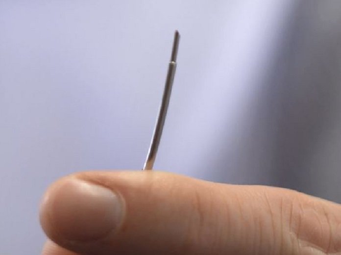
A team of researchers from University College London and Queen Mary University of London has just published details of a groundbreaking cardiology needle that’s capable of imaging the heart’s soft tissue from within. They used the revolutionary all-optical ultrasound imaging system for heart surgery in pigs, successfully capturing high-resolution images while up to 2.5 cm away from the needle tip.
Essentially, the imaging needle works with a built-in miniature optical fiber that transmits brief pulses of light which eventually generate ultrasonic pulses. The ultrasound pulses travel away from the needle, reflecting off soft tissues before being detected by a second optical fiber in the needle housing.
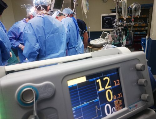
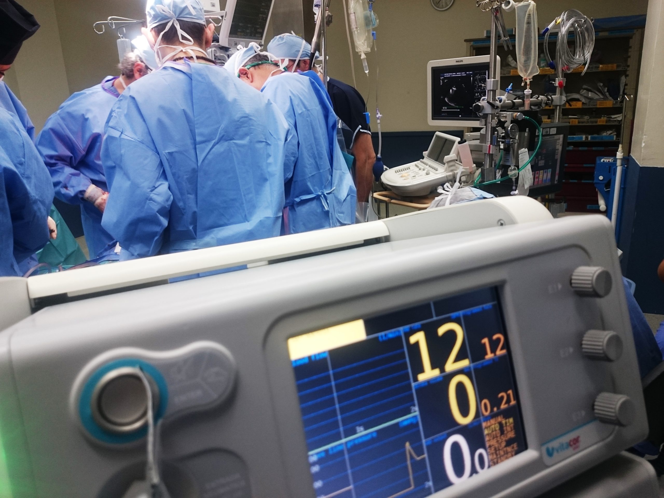
This all-optical ultrasound device does what have never been achieved before — for the first time; live imaging can be captured directly from inside the heart during keyhole surgery. The technology boasts the potential to provide surgeons with significant improvements over current practice. At the moment, surgeons only rely on preoperative imaging combined with external ultrasound probes.
The study co-lead and consultant cardiologists at QMUL and Barts Heart Center, Dr. Malcolm Finlay says, “The optical ultrasound needle is perfect for procedures where there is a small tissue target that is hard to see during keyhole surgery using current methods and missing it could have disastrous consequences.”
The imaging needle produces ultrasound with the help of a novel composite material which consists of a mesh of carbon nanotubes encased in silicone located on the tip of the optical fiber. The pulsed light generated from the fiber is absorbed by the carbon nanotubes and produces an ultrasound wave as a result of the photoacoustic effect. The second innovation supports the detection of reflected ultrasound waves at a very small scale, and it’s all thanks to the highly sensitive optical fibers incorporating polymer optical microresonators.
The exceptional speed of the ultrasound emission and detection process produces novel temporal and spatial resolution of the images. At the moment, the ultrasound system can deliver live imaging with a resolution as fine as 64 microns — this is roughly the width of nine red blood cells. Moreover, the system allows the tracking of the heart walls and valves in real time. The imaging system also has other applications besides cardiology — it can be used in a variety of minimally invasive procedures and also opens up the possibility of futuristic applications including inwomb surgery.
At the moment the team of researchers is looking to translate the system for clinical use in patients.
